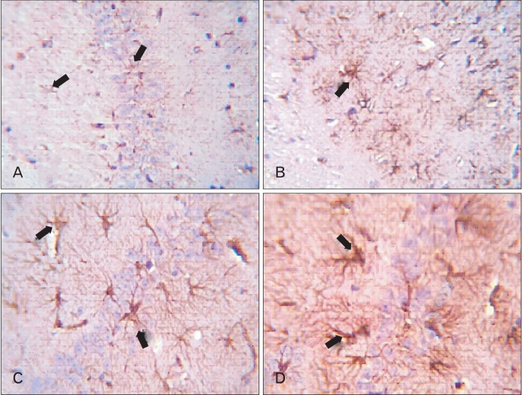
Fig. 8.
CA1 area of hippocampus. (A) Adult balanced diet fed rats shows, slight positive brown glial Fibrillary acidic protein (GFAP) reaction small non branched dispersed astrocytes (arrows). (B) Aged balanced diet fed rats shows, positive brown expression of GFAP in small branched astrocytes (arrow). (C) Adult cholesterol diet fed rats shows, positive brown expression of GFAP in large branched astrocytes (arrows). (D) Aged cholesterol diet fed rats shows, dense positive brown expression of GFAP in large branched astrocytes (arrows) (A-D, GFAP, ×1,000).

