
Figure 5. Lack of preferential innervation between s/dPCs and CCKBCs.
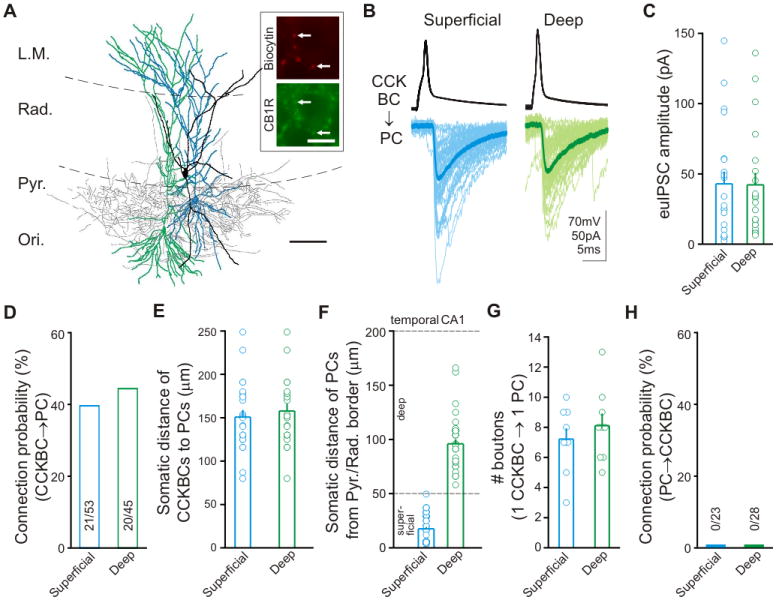
(A) Reconstruction of an sPC (blue), a dPC (green), and a CCKBC (soma and dendrites, black; axons, gray); scale bar: 100μm. Inset: CB1R expression in the CCKBC boutons; scale bar: 10μm. (B) Representative traces from a presynaptic CCKBC and dPC (green) and sPC (blue) in the presence of CB1R antagonist AM251 (10μM) to block the tonic inhibition of GAB A release from CCKBCs (Lee et al., 2010). (C to F) Summary data of euIPSC amplitudes (C), connection probability between CCKBCs to PCs (D), somatic distances between CCKBC-sPC and CCKBC-dPC pairs in the temporal CA1 (E), and somatic locations of the paired recorded s/dPCs with respect to the Pyr/Rad border (F). (G) Number of putative synaptic terminals of single CCKBCs onto single sPCs and dPCs (n=9 for both). (H) Excitatory connection probability from PCs to CCKBCs.