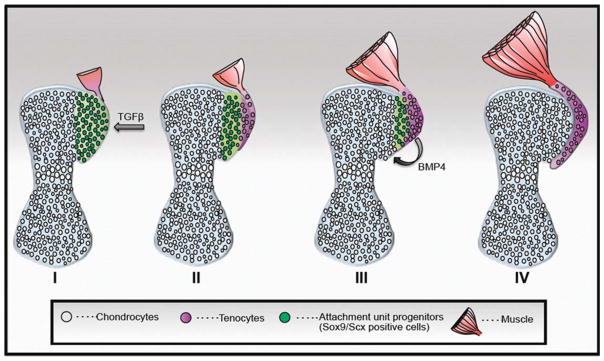
FIGURE 2.

Schematic model for attachment unit formation in situ by segregation of a common progenitor pool to tenocytes and chondrocytes. At the onset (I), the bone anlage comprises differentiated chondrocytes (gray), and the attachment unit domain contains Sox9/Scx-positive progenitors (green). Next (II and III), progenitor cells gradually differentiate to tendon cells from one side (purple) and cartilage cells on the other side (gray) and form the attachment unit (IV). Although specification of attachment unit is regulated by TGFβ signaling (I), their differentiation to chondrocytes is regulated by BMP4 signaling from tendon progenitor cells (Blitz et al., 2013).
