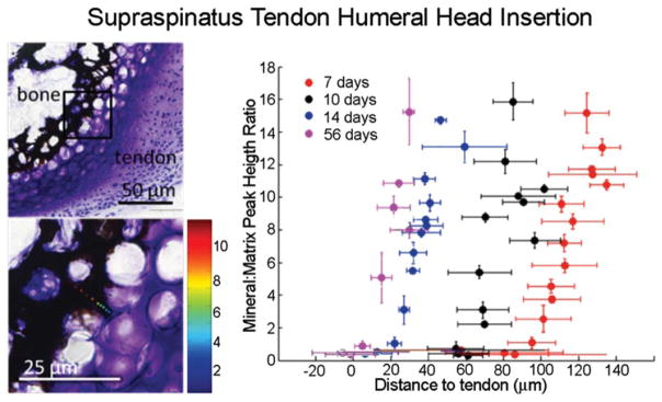
FIGURE 4.

Spatial gradients in mineral (as determined using Raman spectroscopy) form between tendon and bone at the developing entheses from the onset of endochondral ossification (7 days in the mouse supraspinatus tendon enthesis, as shown in the Von Kossa/Toluidine Blue stained sections on the left). Reproduced with permission from Schwartz et al., 2012.
