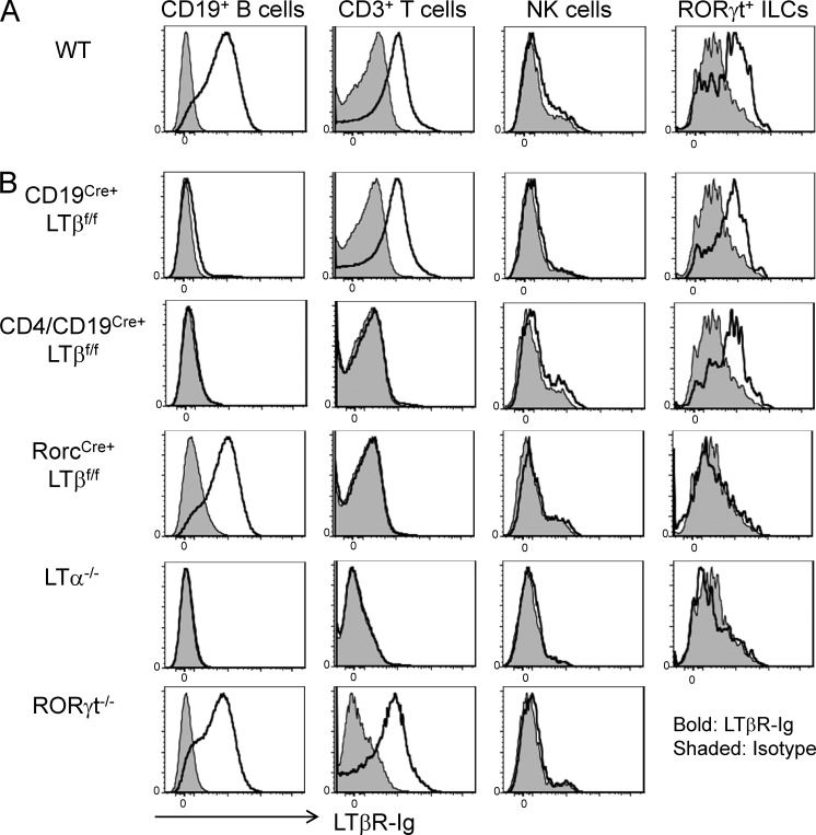
Figure 1.
RORγt+ ILCs but not NK cells express LT. (A and B) Splenocytes from wild-type (A) and CD19Cre+LTβf/f, CD4/CD19Cre+LTβf/f, RorcCre+LTβf/f, LTα−/−, and RORγt−/− (B) mice were stimulated with 20 ng/ml PMA for 20 h. Stimulated cells were then labeled with biotinylated LTβR-Ig for LT staining and assessed by flow cytometry to detect LT expression. Data are representative of two independent experiments.

