Figure 3.
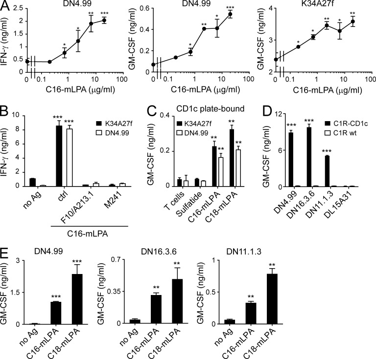
mLPA-specific T cells recognize synthetic mLPA analogues. (A) T cell clones DN4.99 (left and middle) and K34A27f (right) were stimulated with synthetic C16 mLPA presented by purified primary B cells. (B) DN4.99 and K34A27f T cells were stimulated with purified primary B cells unloaded (no Ag) or loaded with 10 µg/ml C16 mLPA in the presence or not (ctrl) of two different anti-CD1c mAbs (F10/A213.1 or M241). (C) The same T cells were either cultured alone (T cells) or stimulated with plastic-bound sCD1c molecules loaded with control lipid (sulfatide) or synthetic mLPA (C16 or C18). (D) Recognition of C1R-CD1c but not C1R WT by CD1c self-reactive T cell clones DN4.99, DN16.3.6, and DN11.1.3, each obtained from a different donor. The CD1c-restricted M. tuberculosis–specific DL15A31 T cell clone was used as a negative control for irrelevant specificity. (E) Activation of T cell clones DN4.99 (left), DN16.3.6 (middle), and DN11.1.3 (right) in the presence of purified primary B cells and synthetic C16 or C18 mLPA (or without antigen, no Ag). T cell activation was evaluated by measurement of IFN-γ and/or GM-CSF release, expressed as mean ± SD. Results are representative of at least three independent experiments. *, P < 0.05; **, P < 0.01; ***, P < 0.001, determined by two-tailed Student’s t test.

