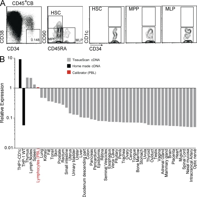
Figure 8.
CD1c expression pattern in human hematopoietic precursors and healthy organs. (A) Normal hematopoietic precursors lack CD1c expression. Cord blood (CB) cells from healthy donors were stained with the indicated mAbs (CD34-FITC, anti–CD45-APCCy7, CD38-PECy7, CD45RA-PB, CD90-APC, and anti–CD1c-PE). The boxes represent the gates used to identify HSCs, multipotent progenitors (MPPs), and multilymphoid progenitors (MLPs), according to Doulatov et al. (2012). Representative dot plots from at least three independent experiments are shown. (B) CD1c mRNA expression in human tissues. RT-qPCR for CD1c expression was performed on cDNAs from human tissues contained in the TissueScan Real-Time Human Major Tissue Panels II (OriGene), according to the manufacturer’s instructions. We added two custom cDNAs, produced by us from THP1-CD1c and THP1 WT cell lines, as positive and negative controls for CD1c expression. The data obtained were normalized against a calibrator signal, provided by the level of CD1c cDNA detection in PBMCs. CD1c expression was detected in lymph nodes, spleen, and thymus, whereas it was almost undetectable in all of the other tissues.

