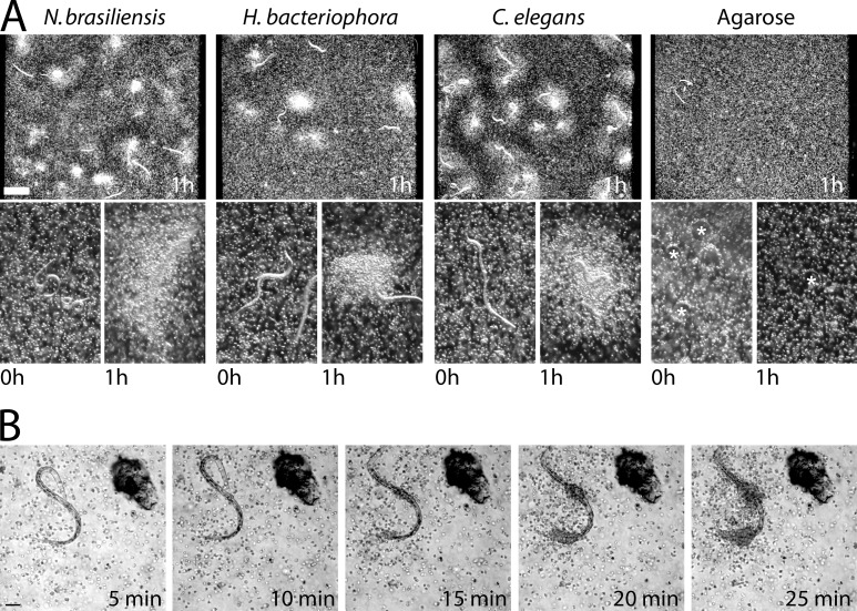
Figure 1.
Eosinophil migration in response to nematodes. (A) Darkfield dissection scope images of bone marrow–derived eosinophils cultured with nematodes. Low-power images (top) show eosinophil accumulation after 1 h. High-power images (bottom) show nematodes before and 1 h after eosinophil accumulation. Agarose beads are marked with asterisks. (B) Images taken from a differential interference contrast time-lapse video (Video 1) of eosinophils migrating toward a C. elegans dauer larva. (A and B) Results represent three independent experiments. Bars: (A) 500 µm; (B) 50 µm.

