Figure 7.
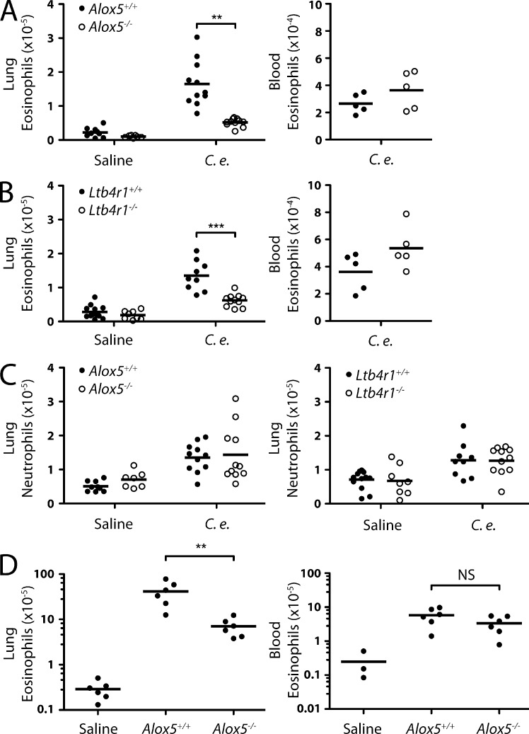
Involvement of leukotrienes in eosinophil accumulation in the lung. (A and B) Flow cytometry analysis of eosinophil accumulation in the lungs (left; three experiments pooled) and blood (right) of Alox5−/− (A) and Ltb4r1−/− (B) mice 24 h after injection of C. elegans dauers or saline. (C) Flow cytometry analysis of neutrophil accumulation in the lungs of Alox5−/−, Ltb4r1−/−, and wild-type mice 24 h after injection of C. elegans dauers or saline. (D) Flow cytometry analysis of eosinophil accumulation in the lungs (left) and blood (right) of Alox5−/− and wild-type mice 9 d after infection with N. brasiliensis larvae. Numbers of eosinophils in uninfected mouse lung (from A) are shown for comparison. (A–D) Results are pooled from three (A–C) or represent two (D) independent experiments. Horizontal bars indicate the mean. **, P < 0.005; ***, P < 0.0005.

