
Figure 2.
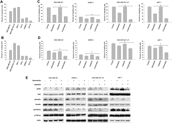
Saracatinib combined with lapatinib efficiently blocks migration and invasion of human breast cancer cell lines. (A) Percentage of migration of MDA-MB-361, MDA-MB-361-LR, JIMT-1, KPL-4, SKBR-3, and BT474, human breast cancer cell lines grown in complete medium measured by wound-healing assay. Results are presented as the percentage of the total distance of the original wound enclosed by cells. (B) Percentage of invasion of MDA-MB-361, MDA-MB-361-LR, JIMT-1, KPL-4, SKBR-3, and BT474 human breast cancer cell lines grown in complete medium, measured by fibroblast monolayer invasion assay. Results are expressed as percentage of monolayer fibroblast invasion. (C) Percentage of migration of MDA-MB-361, SKBR-3, MDA-MB-361-LR, and JIMT-1 treated with saracatinib (0.2 μM), lapatinib (1 μM), or both, measured by wound-healing assay. The results are presented as the percentage of the total distance of the original wound enclosed by cells. (D) Percentage of invasion of MDA-MB-361, SKBR-3, MDA-MB-361-LR, and JIMT-1 treated with saracatinib (0.2 μM), lapatinib (1 μM), or both measured by fibroblast monolayer invasion assay. Results are expressed as percentage of invasion of fibroblast monolayer. (E) Western blot analysis of total cell lysates from MDA-MB-361, SKBR-3, MDA-MB-361-LR, and JIMT-1 treated with saracatinib (0.2 μM), lapatinib (1 μM), or both. The data represent the mean (±standard deviation, SD) of three independent experiments, each performed in triplicate, and are presented relative to control (cells treated with DMSO). Error bars indicate SDs. Asterisks indicate statistical significance of combined treatment versus lapatinib alone, determined with Student t test (*two-sided P < 0.5 × 10⁻¹; **two-sided P < 0.5 × 10⁻²). The relative optical density of phosphoprotein levels normalized to the actin level is shown.