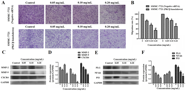
Figure 6. Dose-response study of ESWE on the migration of cells transfected with siRNA of PKCβ and normal SMMC-7721 cell in vitro.
(A) Photographs of the knockdown cell and wide type cell migration through the polycarbonate membrane stained by 0.2% crystal violet. (B) Quantification of the number of cells (transfected with siRNAs of PKCβ and wide type cells) migrating through the polycarbonate membrane. Data represents the means ± SD from three repeated experiments. Five wells were treated in each experiment. (C) Bands were corresponding to MMP-2, MMP-9, CXCR4 and GAPDH in SMMC-7721 cells. Full-length blots are presented in Supplementary Figure 6C. (D) Results were quantified by densitometry analysis of the bands form (C) and then normalization to GAPDH protein. (E) Bands were corresponding to PLG, NF-kb, P53 and GAPDH in SMMC-7721 cells. Full-length blots are presented in Supplementary Figure 6E. (F) Results were quantified by densitometry analysis of the bands form (E) and then normalization to GAPDH protein. Quantitation data showed ESWE decreased the proteins levels involved in cell migration in a dose-dependent manner compared to the untreated control. Values are expressed as means ± SD (n = 3). * p < 0.05, ** p < 0.01, vs. control.

