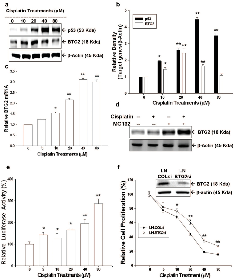
Figure 2. Cisplatin modulates p53 and BTG2 expression in LNCaP cells and BTG2 mediates partial cisplatin-induced growth inhibition in LNCaP cells.
(a) LNCaP cells were treated with various concentrations of cisplatin, as indicated, for 24 hours. Cells were lysed and expressions of BTG2 and p53 were determined by immunoblotting assay.(Cropped gel) (b). Data of quantitative immunoblot analysis are expressed as relative density (mean ± SE; n = 3). (c) LNCaP cells were treated with various concentrations of cisplatin, as indicated, for 24 hours. BTG2 mRNA levels were determined by RT-qPCR. (d) LNCaP cells were pretreated with or without MG132 for 2 hours and then treated with 80 μM cisplatin for 24 hours. Cells were lysed and expression of BTG2 was determined by immnuoblotting assay.(Cropped gel) (e) Luciferase activity of BTG2 reporter vectors-transfected LNCaP cells after treated with various concentrations of cisplatin for 24 hours. (f) LN-BTGsi cells (BTG2 knockdown LNCaP cells) and LN-COLsi cells (mock-knockdown LNCaP cells) were treated with indicated concentrations of cisplatin for 48 hours. Cell proliferation was measured by the CyQUANT cell proliferation assay. Each value is presented as the % in relation to the control (solvent-treated) group. Data are shown as the mean percentage ± SE (n = 6) (*P < 0.05, **P < 0.01).

