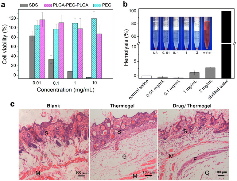
Figure 3. Biocompatibility evaluation of the triblock copolymer PLGA-PEG-PLGA synthesized in this study.
(a) Cytotoxicity of PLGA-PEG-PLGA against the MC3T3 cell line after incubating for 24 h with PEG of number average MW Mn ~ 2000 Da (PEG2000) as the negative control and SDS as the positive control (n = 4). The absorption in the CCK-8 test of the untreated sample (culture medium with 10% fetal bovine serum) was defined as 100%. (b) Hemolysis of rabbit blood cells after being suspended in PLGA-PEG-PLGA aqueous solutions with normal saline as the medium at 37°C for 1 h (n = 3). Complete hemolysis occurred in the group of distilled water, which was defined as 100%. The inserted image shows the medium color of the hemolysis of red blood cells after centrifugation. (c) Optical micrographs of hematoxylin-eosin (HE)-stained slices of surrounding tissues 7 days after subcutaneous injection of the indicated solutions. S: skin tissue; M: muscle tissue; F: fibrous tissue; G: thermogel.

