
FIGURE 4.
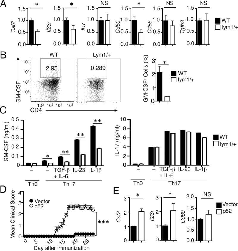
Regulation of GM-CSF induction and encephalitogenicity of T cells. (A) qRT-PCR analysis of relative mRNA levels (normalized to the control Actb) of the indicated genes in CD4+ T cells freshly isolated from WT and lym1/+ mice immunized with MOG35–55 for 7 days. (B) CNS CD4+ T cells from EAE-induced WT and lym1/+ mice (day 14 after immunization) were stimulated for 4 hours with PMA plus ionomycin and subjected to ICS and flow cytometry to determine the frequency of GM-CSF producing cells. Data are presented as a representative flow cytometry graph (left) and mean value (right). (C) Naive CD4+ T cells isolated from WT and lym1/+ mice were stimulated for 72 hours with plate-bound anti-CD3 and anti-CD28 under Th0 or Th17 conditions. Differentiated Th17 cells were rested for 2 days, followed by the second stimulation of 72 h with anti-CD3 and anti-CD28 in the absence (−) or presence of the indicated cytokines. ELISAs were performed for GM-CSF and IL-17 produced. (D) lym1/+ CD4+ T cells were infected with vector or p52. After 48h, cells were adoptively transferred into Rag1−/− mice (n=4). The recipient mice were immunized for EAE induction. (E) qRT-PCR analysis of the indicated genes in CD4+ T cells isolated from mice of (D). Data are representative of two or more independent experiments.