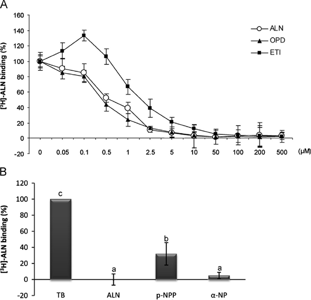
Fig. 1.
Displacement of osteoblast [3H]-ALN binding by other BPs and protein phosphatase substrates. (A) ROS 18/2.8 osteoblastic cells were incubated with 30 nM [3 H]-ALN in the presence of different concentrations (0–500 µM) of unlabeled BPs: ETI (▪), OPD (▲) and ALN (o). Each value is the mean±SD of results from three separate experiments performed in triplicate. B) ROS 17/2.8 cells were incubated with 30 nM [3H]-ALN in the absence (total binding, TB) or presence of 200 µM unlabeled ALN or 8 mM of the phosphatase substrates p-nitro-phenylphosphate (p-NPP) or α-naftylphosphate (α-NP). Results expressed as a percent of total binding are the average±SD. Data were analyzed by one-way analysis of variance, and the Bonferroni test was used for mean comparison. Same letters indicate p>0.05, whereas different letters indicate p<0.05.

