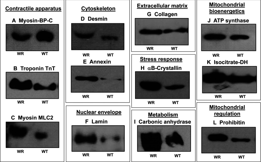
Figure 4. Immunoblot survey of proteins with a changed abundance in WR muscle as revealed by proteomics.
Shown are representative immunoblots with expanded views of immuno-decorated bands labelled with antibodies to MBP-C (A), troponin subunit TnT (B), myosin light chain MLC2 (C), desmin (D), annexin (E), lamin (F), collagen (G), αB-crystallin (HSPB5) (H), carbonic anhydrase isoform CA3 (I), mitochondrial ATP synthase (J), isocitrate dehydrogenase (K) and prohibitin (L). Lanes 1 and 2 represent WR versus WT muscle preparations, respectively.

