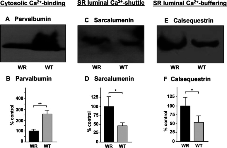
Figure 6. Immunoblot analysis of Ca2+-binding proteins with a changed abundance in WR muscle.
Shown are representative immunoblots with expanded views of immuno-decorated bands labelled with antibodies to the cytosolic Ca2+-binding protein parvalbumin (A, B), the luminal Ca2+-shuttle protein sarcalumenin of the SR (sarcoplasmic reticulum) (C, D) and the luminal Ca2+-binding protein calsequestrin of the terminal cisternae region of the SR (E, F). Lanes 1 and 2 represent WR versus WT muscle preparations, respectively. The comparative immunoblot analysis was statistically evaluated using an unpaired Student's t test (n=5; *P<0.05; **P<0.01).

