
Fig. 4.
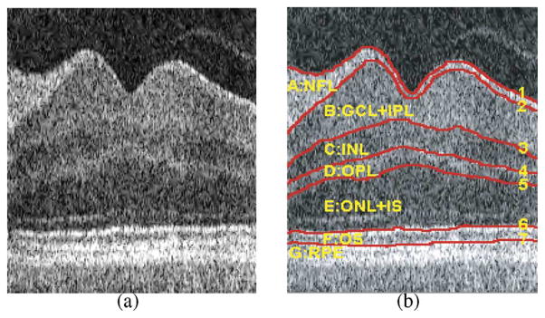
Intraretinal layers in 3-D OCT images. (a) A 2-D slice from the center of a volumetric OCT image—the dip corresponds to the fovea. (b) Seven surfaces (labeled 1–7) and corresponding retinal layers (NFL: nerve fiber layer; GCL+IPL: ganglion cell layer and inner plexiform layer; INL: inner nuclear layer; OPL: outer plexiform layer; ONL+IS: outer nuclear layer and photoreceptor inner segments; OS: photoreceptor outer segments and RPE: retinal pigment epithelium).