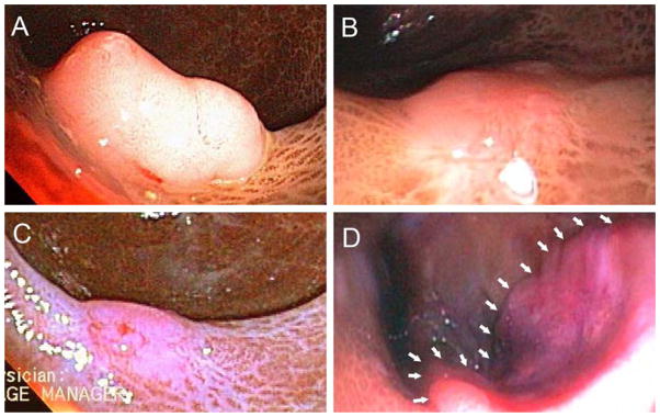
Figure 1. Endoscopic Findings.

Panel A shows our patient’s rectal polyp prior to initial polypectomy. Panel B displays the site six-months later when additional polypectomy revealed residual adenoma. Panel C shows a polypectomy scar after an additional three months. Multiple biopsies obtained at this time showed no adenomatous tissue. Panel D demonstrates the rectal adenocarcinoma found 28 months after the unremarkable sigmoidoscopy with biopsy (white arrows outline the proximal tumor margin).
