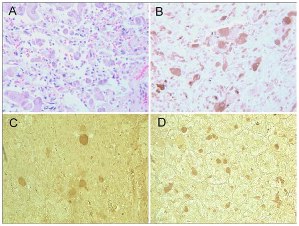
Figure 2. Histopathological and Immunohistochemical Evaluation of Pheochromocytoma.
Histopathological evaluation of the pheochromocytoma showed connective tissue with clusters and isolated pleomorphic tumor cells with abundant and granular cytoplasm (Panel A, H&E stain). Immunohistochemical staining for choline acetyltransferase (ChAT) shows strong cytoplasmic staining of most tumor cells in this field (Panel B, anti-ChAT stain). Two of five additional archival specimens stained positively for ChAT (Panels C & D, anti-ChAT stain). ChAT = choline acetyltransferase.

