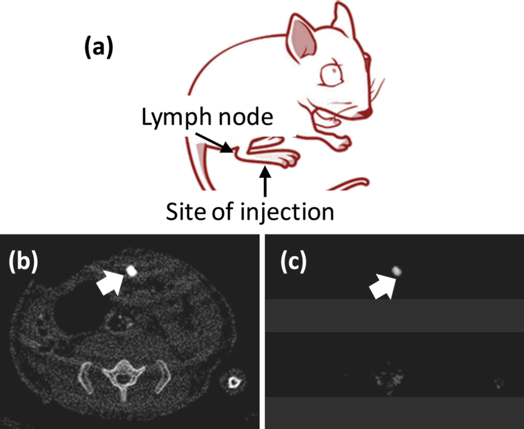
Figure 8.
In vivo noninvasive spectral CT imaging of sentinel lymph nodes. (a) A cartoon illustrating the site of injection and the area of interest. 150 µL of nanobeacons were injected intradermally in all the cases; (b) regional sentinel lymph nodes were clearly contrasted in conventional CT; (c) K-edge contrast of accumulated gold in the lymph node was selectively imaged with spectral CT.

