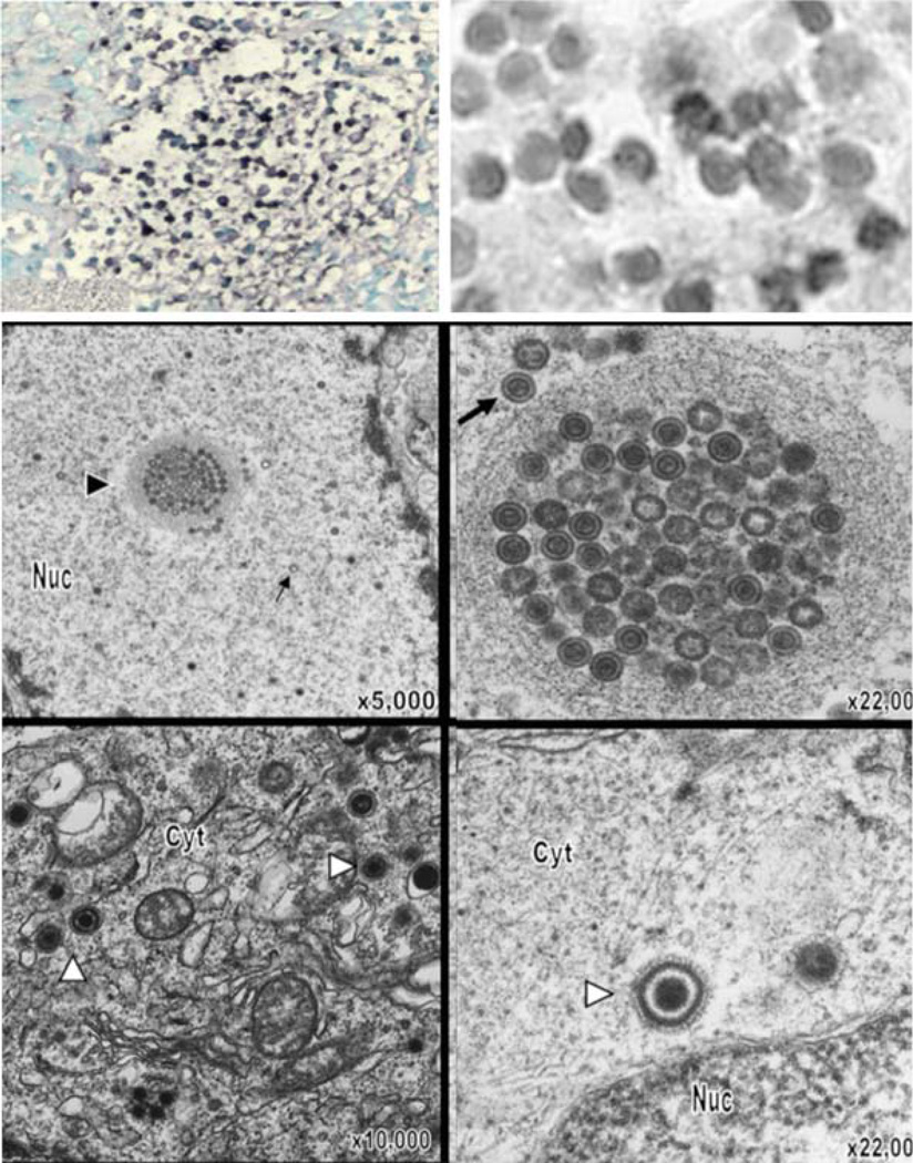
Fig. 1.
VZV infection of T cells in thymus/liver (T cell) xenografts in the SCID mouse model. On day 7 after infection, infected T cell xenografts were tested for VZV DNA by in situ hybridization; darkly stained cells indicate VZV DNA in T cells visualized at lower (left) and higher magnification (right; ×786). By electron microscopy on day 14 after infection, VZV nucleocapsids were present in the nuclei with most containing a dark VZV DNA core (large arrows); some empty capsids were also seen (small arrows). Virions were abundant in T cell nuclei, both individually and in clusters (arrowhead), also shown at higher magnification. Complete virions were found in the cytoplasm. Magnification and locations of cytoplasm (cyt) and nucleus (nuc) are as indicated. From Moffat et al. 1995 and Schaap et al. 2005; reproduced with permission

