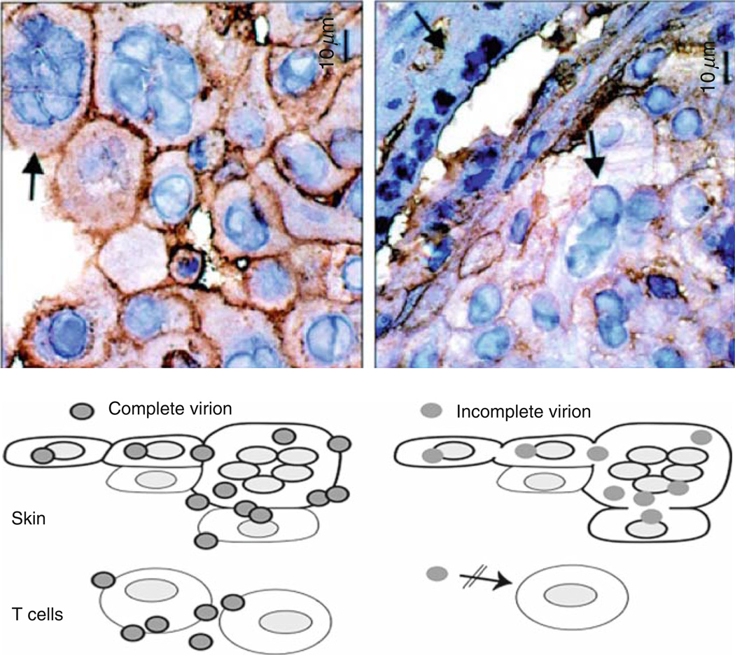
Fig. 2.
Role of cell fusion and polykaryocyte formation in VZV infection of skin xenografts. The upper panels show virion formation in skin xenografts infected with VZV (left panel) and kinase-defective ORF47 mutant (right panel) and examined at day 21 by staining with antibody to VZV gE and hematoxylin. Transmission microscopy demonstrated many intact viral particles in rOkainfected skin cells. Viral particles were incomplete and fewer in rOka47D-N infected skin cells. Arrows indicate polykaryocytes with dark staining of gE in plasma membranes of rOka-infected cells and persistent formation of polykaryocytes in skin infected with the ORF47 kinase mutant despite minimal gE localization to plasma membranes. As illustrated, virus-induced cell fusion, with the characteristic formation of polykaryocytes, supports VZV infection in skin but VZV infection does induce cell fusion. ORF47 kinase mutants do not infect T cells, suggesting that VZV T cell tropism depends upon efficient assembly and release of complete virions. (Adapted from Besser et al. 2004; figures reproduced with permission)

