
Figure 3.
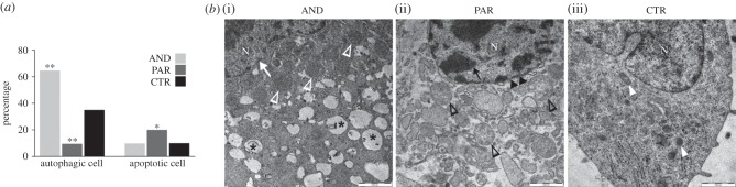
Divergent subcellular features of AND and PAR placentae at day 20 of sheep development. (a) Transmission electron microscopy analysis revealed that AND placentae were characterized by a very high number of autophagic cells (22/34 versus 10/28; **p = 0.0029), while PAR placentae exhibited a twofold decrease (**p = 0.0021) of autophagic cells and an increased number of apoptotic cells (8/32 versus 2/28; *p = 0.0324). (b) Electron micrograph of autophagic, apoptotic and normal cells from AND, PAR and biparental control placentae, respectively. (i) The AND cell had an intact nuclear membrane (white arrow), numerous autophagosomes (asterisk) and swollen mitochondria (white arrowheads). (ii) The PAR cell exhibited fragmented cytoplasm, a high number of apoptotic bodies (black empty arrowheads) and condensed chromatin (black arrow), and its nuclear membrane (black double arrowheads) lacked integrity. (iii) Control biparental cell (mitochondria: white arrowheads). N, cell nucleus. Scale bar, 1000 nm.