Figure 4.
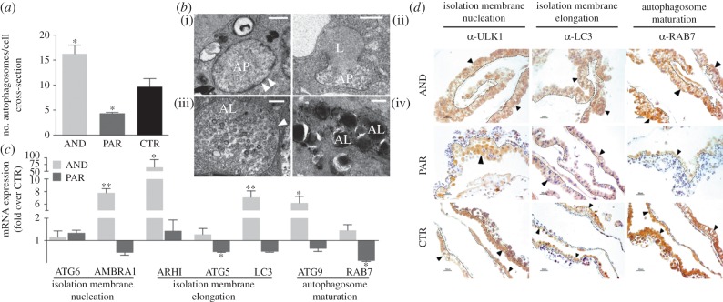
Increased level of autophagy in AND placentae. (a) An increased number of autophagosomes (*p < 0.05) in AND placentae and a decreased number of autophagosomes (*p < 0.05) in PAR placentae indicates the deregulation of autophagy in both uniparental models. To study whether this deregulation was due to an acceleration or blockage of autophagy at any particular stage, ultrastuctural (b) and molecular (c) analyses were performed. (b) The presence of all stages indicative of autophagy: (i) autophagosome—AP (white arrowheads indicate double membrane); (ii) fusion of autophagosome with lysosome—L; (iii) autophagolysosome—AL (white arrowheads indicate single vacuolar membrane); and (iv) mature autophagolysosome—AL, indicated that this process was not blocked at any particular stage in AND placentae. (c) Upregulated expression (more than fivefold over CTR, **p < 0.01; *p < 0.05) of genes regulating autophagy, from the initial isolation membrane nucleation and elongation to the complete autophagosome formation and maturation in AND placentae. (d) AND placentae also exhibited a higher protein expression of autophagosome markers (ULK1, LC3 and RAB7). Trophoblastic cells (trophoblast layer shown by arrows and delimited by dashed lines) were intensely stained in AND placentae, while less intense or absent signals of respective markers were observed in PAR placentae.

