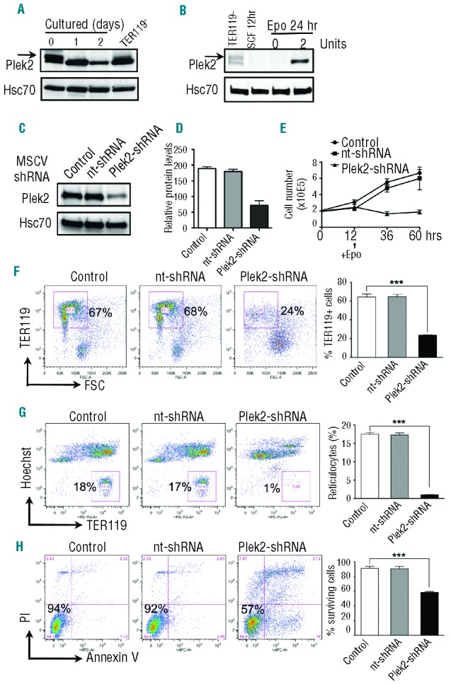
Figure 2.

Plek2 is required for the early stage of terminal erythropoiesis. (A) Western blot analysis of plek2 on different days of cultured mouse fetal liver erythroblasts in Epo medium together with TER119 positive fetal liver cells. Hsc70 was used as a loading control. (B) Western blot analysis of plek2 on freshly purified TER119 negative fetal liver erythroblasts, cells cultured in SCF medium for 12 h, and cells cultured in SCF medium for 12 h followed by 24-h Epo medium culture. Hsc70 was used as a loading control. (C) TER119 negative fetal liver erythroblasts were transduced with retroviruses encoding the indicated shRNAs and cultured in SCF medium for 12 h. The cells were changed to Epo medium and continued to culture for 12 h followed by Western blot analysis of plek2. Nt-shRNA represents a non-targeting shRNA. Hsc70 was used as a loading control. (D) Quantification of (C). Data were obtained from 3 independent experiments. (E) Cell growth and quantity analysis after knockdown by indicated shRNAs. Same as C except that the cells were cultured for two more days after change to Epo medium. The cells were counted using a hemocytometer on each day. (F) Flow cytometric analysis of TER119 level in the indicated cells from (E). Right bar graph is the statistical analysis using Student’s t-test. *** P<0.001. (G–H) Analysis of mouse fetal erythroblast enucleation and apoptosis during in vitro culture. Same as (E), the cells after culture were analyzed by flow cytometric analysis for enucleation (G) and apoptosis (H) using Hoechst 33342 and TER119, and propidium iodide (PI) and Annexin V, respectively. Statistical analysis using Student’s t-test is on the right. ***P<0.001.
