
Figure 2.
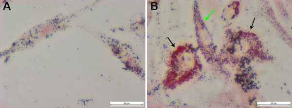
Oil Red O staining of human primary cultured VSMCs. (A) Oil Red O staining of normal human carotid VSMCs after 7 weeks in the 3rd passage. (B) Oil Red O staining of human plaque-derived VSMCs with two distinctly different morphologies after 7 weeks in the 3rd passage. The big, flattened shape ones (black arrow) contain much more lipid than the fusiform ones (green arrow).