
Figure 3.
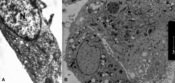
Transmission electron micrograph of human plaque-derived VSMCs cultured for 7 weeks in the 3rd passage. (A) The ultrastructure of fusiform plaque-derived VSMCs. Arrows show myofilaments with dense bodies. ×6000. (B) The ultrastructure of human plaque-derived VSMCs with a big, flattened shape. N: nucleus; M: mitochondria; RER: rough endoplasmic reticulum; LD: lipid droplets.