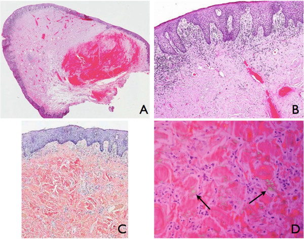
Figure 2.

Histological and immunohistochemical tests. (A) Panoramic section (haematoxylin and eosin: ×0.6). (B) Irregular acanthosis, lymphocytic infiltration and amyloid deposits (haematoxylin and eosin: ×4). (C) Congo red stain of amyloid (×10). (D) Congo red stain showing characteristic apple-green birefringence (arrows) under polarized light (×60).
