Figure 2.

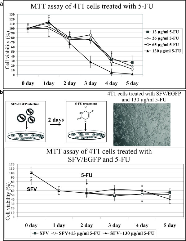
Evaluation of 4 T1 cell proliferation after 5-FU treatment and in combination with SFV infection. (a) 5-FU treatment. 4 T1 cells were grown in cell culture medium (24-well plates) containing the indicated concentrations of 5-FU. The MTT cell viability assay was performed every day for 5 days. The diagram shows the cytotoxic effect of 5-FU on 4 T1 cells as the percentage of viable cells relative to the control (untreated cells). (b) Schematic representation of the combined treatment with SFV and 5-FU. The cells were infected with SFV/EGFP particles, and the medium was replaced 2 days later with medium containing 5-FU. The MTT cell viability assay was performed every day for 5 days. The arrows designate the day of infection (SFV) and the beginning of the drug treatment (5-FU). The diagram shows the cytotoxic effect of 5-FU following SFV infection as the percentage of viable cells relative to the control (untreated cells). The error bars indicate the standard error of 3 independent experiments. The microscopy image shows a 4 T1 cell monolayer at day 5 after treatment with SFV and the highest concentration of 5-FU.
