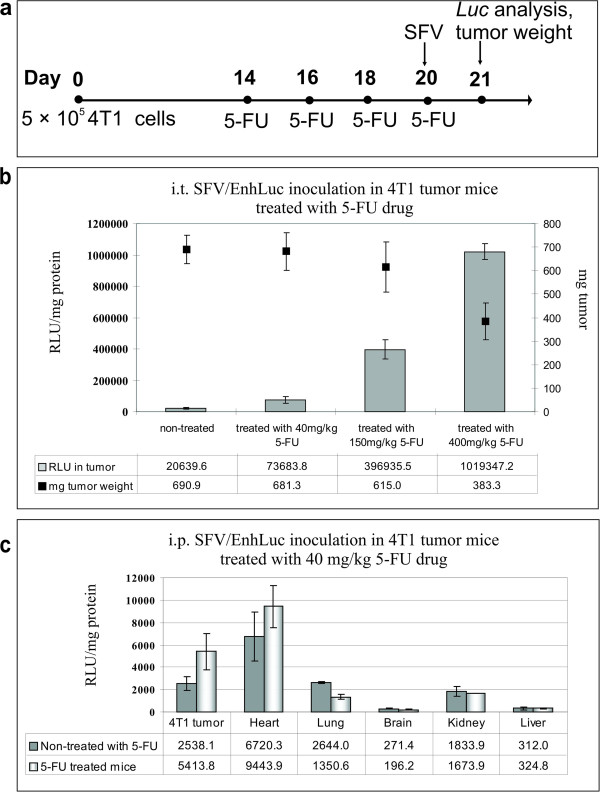
Figure 5.

SFV expression in 4 T1 tumor-bearing mice treated with 5-FU. (a) Experiment design: Balb/c mice (n = 5 in each group) were subcutaneously inoculated with 4 T1 cells; beginning on day 14, the mice were treated four times with 5-FU, every other day (40 mg kg−1, 150 mg kg−1 or 400 mg kg−1). On day 20, after the last 5-FU administration, the mice were i.t. or i.p. inoculated with SFV/EnhLuc virus particles. Tumor weight and Luc gene expression were measured 24 h after viral inoculation. (b) Intratumoral Luc gene expression after i.t injection of SFV/EnhLuc virus particles in 5-FU-treated mice. Luciferase activity was measured in tumor homogenates 24 h after virus inoculation. Tumor weights were measured prior to homogenization (scale on the right). (c) SFV/EnhLuc virus biodistribution in 4 T1 tumor-bearing mice treated with 40 mg kg−1 5-FU. Luciferase activity was measured in tumor and organ homogenates 24 h after i.p. virus inoculation. The graphs present the RLUs per mg protein in each organ or tumor (see Methods section). The results are presented as the means ± s.e. The average RLU values and tumor weights are indicated in the tables. RLU, relative light unit.
