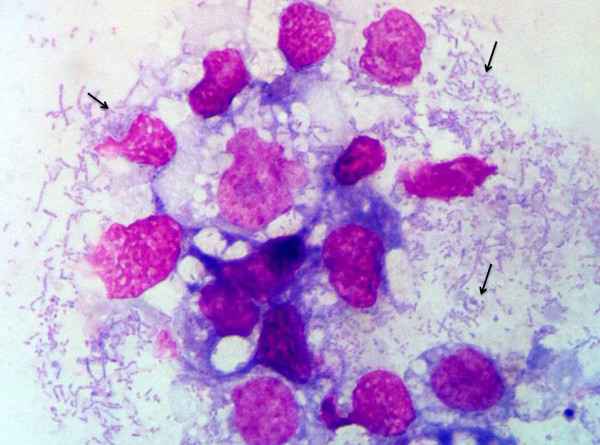
Figure 3.

ISE-6 cells at 6 weeks post inoculation. Photomicrograph of cytocentrifugation preparation of infected ISE-6 cells. The cells contain abundant, pleomorphic, rod-shaped bacteria (Arrows). (Wright-Giemsa stain, 100X magnification) Images were captured with Mioticam 580 with 5.0 megapixel (Motic North America, British Columbia, Canada).
