Fig. 2.
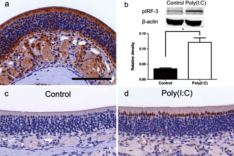
Expression of TLR3, phospho-IRF-3 (Ser 396) and phospho-NF-κBp65 (Ser 276) in the mouse olfactory mucosa. a The expression of TLR3 was observed mainly in the apical part of the supporting cells and in the cytoplasm of the acinar cells of Bowman’s glands. b Western blot image showing phospho-IRF-3 expression. Bar graph represents the relative density of each band normalized to β-actin as internal control. It was significantly greater in the Poly(I:C) group than in the control group (n = 3, p < 0.005). c In the control group, immunolabeling for phospho-NF-κBp65 was observed very weakly in the nuclei of some supporting cells. d In the Poly(I:C) group, immunolabeling for phospho-NF-κBp65 was detected intensely in the nuclei of many supporting cells and acinar cells of Bowman’s glands at 8 h. Bar 150 μm

