
Fig. 1.
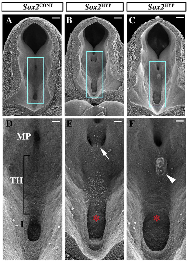
Scanning electron analyses of the posterior ventral diencephalon in E10.5 Sox2CONT and Sox2HYP embryos. (A–C) Posterior-facing views of Sox2CONT (A) and Sox2HYP (B and C) E10.5 diencephala. Scale bars for A–C: 100 μm. (D–F) Magnified images of the regions outlined in blue in (A-C) showing the mammillary region, tuberal hypothalamus, and infundibulum of Sox2CONT (D) and Sox2HYP (E and F) embryos. Sox2CONT mammillary pouches appear as singular midline invaginations. (D) In contrast, Sox2HYP mammillary pouches are often divided by ectopic, undescended tissue (arrow in E). The infundibula of Sox2HYP embryos are expanded relative to those of Sox2CONT embryos (D vs. E, F, red asterisks). A subset of Sox2HYP embryos exhibit a protuberance in the tuberal hypothalamus by E10.5 (arrowhead). Scale bars for D–F: 50 μm. MP: mammillary pouch; TH: tuberal hypothalamus; I: infundibulum.