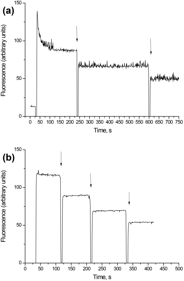
Figure 5.
(a) Membrane potential of the synaptosomes after the addition of D-mannose-coated γ-Fe2O3 nanoparticles. The suspension of the synaptosomes was equilibrated with rhodamine 6G (0.5 µM); when the steady level of the dye fluorescence had been reached, nanoparticles (250 µg/mL) were added (arrows). (b) Rhodamine 6G fluorescence after the addition of D-mannose-coated γ-Fe2O3 nanoparticles (without synaptosomes). Each trace represents four experiments performed with different preparations.

