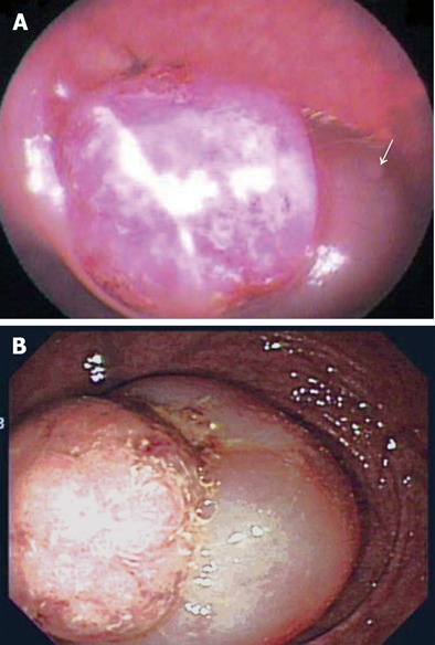
Figure 1.

Endoscopic findings. A: Flexible sigmoidoscopy at a local clinic showing the mass lesion 15 cm from the anal verge, and the partial downward displacement of involved bowel (arrow); B: Colonoscopy at our clinic showing the invaginated bowel with a round mass lesion about 3 cm from the anal verge.
