
Figure 1.
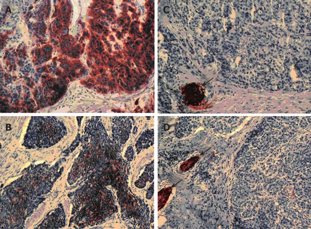
L1 expression in pancreatic neuroendocrine tumours or carcinomas. Immunohistochemical staining was performed by peroxidase method using monoclonal antibody UJ.127 against L1. Poorly-differentiated L1-positive pancreatic neuroendocrine carcinomas (grade 2; A and B) were shown in comparison to well-differentiated L1-negative tumours (grade 1a; C and D). Peripheral nerves (arrows) stained in (C, D) served as internal positive controls (Magnification ×200 (A and C) and ×400 (B and C)).