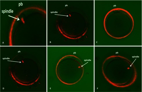
Figure 1.

Birefringent spindles in living human oocytes imaged with the Polscope just before ICSI. A, Birefringent spindles at telophase I. B, Oocytes had visible spindles at metaphase II. C, Oocytes had no visible spindles at metaphase II. D, Oocytes with the spindle located between 0° and 30° relative to the polar body. E, Oocytes with the spindle located between 30° and 60° relative to the polar body. F, Oocytes with the spindle located between 60° and 90° relative to the polar body.
