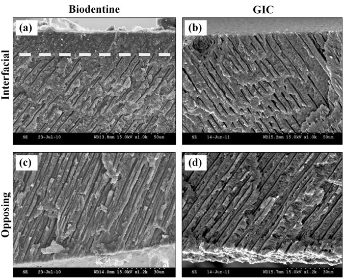
Figure 3.
SEM micrographs of the fractured dentin discs which were exposed to Biodentine (a) or GIC (b). A band of structurally altered dentin extends along the interface, as can be seen by the obliterated dentin tubules above the dotted line in (a), when compared with dentin of the opposite surface of the disc, where no cement was applied (c). (b) No structural changes can be detected in the dentin beneath GIC when compared with dentin of the opposite surface (d).

