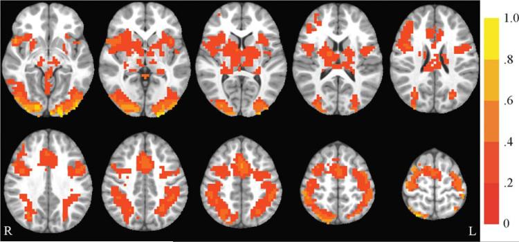
FIGURE 4.
Incongruent vs. fixation activation at baseline. Axial slices (top left z = –8 through bottom right z = 64, spacing = 8 mm) displaying significant activation for the incongruent vs. fixation contrast across all participants at baseline. The background anatomical image is the pediatric template that was used during alignment and is shown using radiological convention. Scale indicates percent signal change.

