
Figure 1.
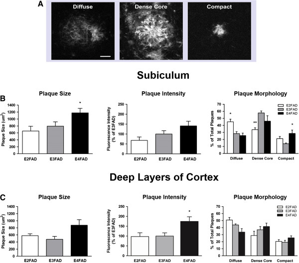
APOE genotype affects Aβ plaque morphology in EFAD subiculum and deep layers of cortex. Morphologically distinct Aβ plaques were visualized in the subiculum and deep layers of the cortex of 6-month-old EFAD mice using immunofluorescence staining with Aβ antibody MOAB2. (A) Heterogeneous plaque types were evident in EFAD brains and grouped into three categories: diffuse, dense core and compact. Scale bar: 12 μm. (B) Subiculum. Left: E4FAD mice exhibited larger plaques on average than E3FAD mice. Center: Fluorescence intensity of plaques did not differ by APOE genotype. Right: E2FAD mice exhibited a greater percentage of diffuse plaques than E3FAD or E4FAD mice, and a lower percentage of dense core plaques than E3FAD mice. Interestingly, E4FAD mice exhibited an increased percentage of compact plaques in the subiculum. (C) Deep layers of the cortex. Left: Plaque size did not differ by APOE genotype. Center: Fluorescence intensity of deep cortical plaques was highest in E4FAD mice. Right: E2FAD mice had a greater percentage of diffuse plaques in this region than E4FAD mice. No differences were detected between APOE genotype for dense core or compact plaques. One-way ANOVA, * P < 0.05. Two-way ANOVA, * P < 0.05, ** P < 0.01.