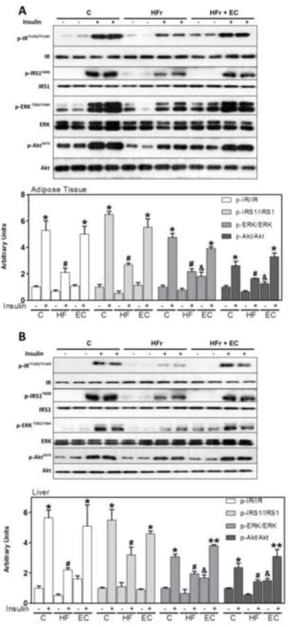
Figure 2. EC supplementation enhances insulin signaling in epididymal adipose and liver tissues in HFr-fed rats.

After 8 weeks on the corresponding diets, rats were fasted overnight then injected with saline or insulin (10 mU/g body weight) and then sacrificed after 10 minutes. Phosphorylation of IR, IRS1, ERK1/2, and Akt are shown for A- epididymal adipose tissue and B-liver. Bands were quantified and results for the HFr and HFr + EC were referred to control group values (C). Results are expressed as the ratio of phosphorylated/total protein level. Results are shown as mean ± SEM of 5 animals/treatment. *, # are significantly different between them and from the insulin untreated groups, &are significantly different from the insulin untreated control and HFr groups, and ** are significantly different from all other groups (p<0.05, one way ANOVA test).
