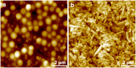
Figure 5.

AFM characterization of Latex 2 and PCC substrates exposed to S. aureus for 5 minutes. Topographical images (10 μm × 10 μm) of a) Latex 2 and b) PCC sample exposed to S. aureus for 5 min followed by exposure to TSB for 18 h. The Z ranges are a) 799 nm and b) 1004 nm and scale bars 2 μm.
