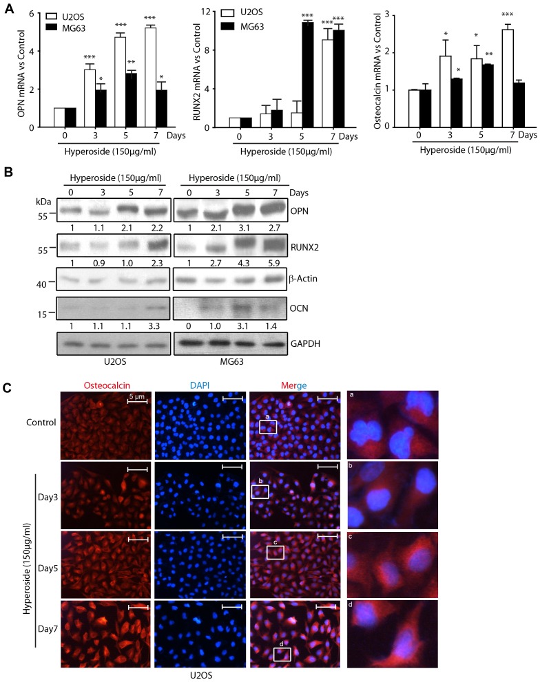
Figure 3. The differentiation is occurred after osteosarcoma cells treating with Hyperoside.
(a) U2OS and MG63 cells were treated with 150 µg/ml hyperoside for 0–7 days. Total RNA were extracted and subjected for the amplification of the indicated transcripts by Real Time PCR. Expression levels were normalized to GAPDH. (b) U2OS and MG63 cells were treated with 150 µg/ml hyperoside for 0–7 days. The whole cell lysates were obtained and fractionated on a 10% SDS-PAGE gel. Next, an immuneblotting assay with anti-OPN, anti-RUNX2, anti-osteocalcin (OCN) or anti-β-Actin antibodies was performed to determine the levels of OPN, RUNX2 and osteocalcin. The numbers below the bands indicated the density of each WB band. (c) U2OS cells were treated with hyperoside for the indicated times. Immunofluorescence analysis of osteocalcin (red) expression was performed. DNA was stained with a fluorescent dye, DAPI.

