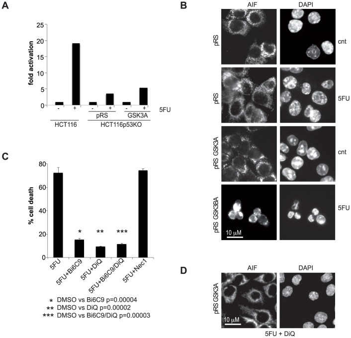
Figure 5. p53-null, GSK3B-silenced colon carcinoma cells treated with 5FU die by RIP1-independent necroptosis.
A) caspase-3/7 activation in HCT116p53KO-pRS and -pRSGSK3A cells treated with 200 µM 5FU for 72 hrs. HCT116 were used as control. Values indicate the fold increase of enzymatic activity of treated cells relative to the untreated cells arbitrarily set as 1. A representative experiment is shown. B) HCT116p53KO-pRS and -pRSGSK3A treated for 30 hrs with 200 µM 5FU and stained with anti-AIF antibody as well as DAPI. C) percentage of cell death of HCT116p53KO-pRSGSK3A after 72 hs treatment with 200 µM 5FU in presence of Bid inhibitor (20 µM Bi6C9), PARP1 inhibitor (100 µM DiQ), Bi6C9+DiQ or Necrostatin-1 (20 µM Nec1). D) HCT116p53KO-pRSGSK3A treated for 30 hrs with 200 µM 5FU in presence of 100 µM DiQ and stained with anti-AIF antibody as well as DAPI.

