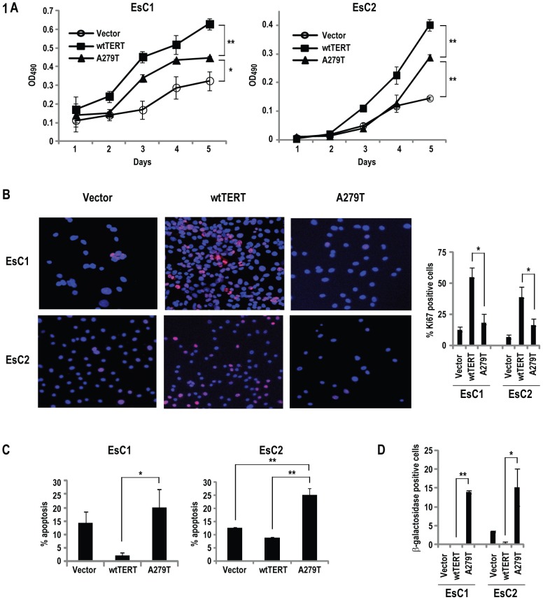
Figure 1. A279T inhibits proliferation of esophageal cancer cells.
(*p<0.05; **p<0.01). A. MTS assay demonstrating inhibition of EsC1 (left) and EsC2 (right) proliferation by A279T relative to wtTERT. B. Immunofluorescence analysis (left panel) with corresponding summary (right panel) of Ki67 expression in esophageal cancer cells expressing wtTERT or A279T (Red: Ki67; blue: DAPI). EsC1 and EsC2 cells expressing A279T exhibit decreased Ki67 levels relative to respective cells expressing wtTERT. C. Annexin V-FITC assay demonstrating A279T-induces apoptosis in EsC2 but not EsC1 cells. Results are expressing as mean ± SD of triplicate experiments. D. Graphic summarization of immunofluorescence analysis of β-galactosidase expression in EsC1 and EsC2 following constitutive expression of wtTERT or A279T. Red: β-galactosidase; blue: DAPI.

