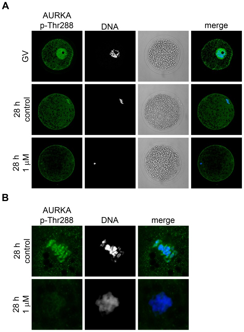
Figure 3. Detection of AURKA (Thr288) phosphorylation.
(A) Fluorescent images of AURKA p-Thr288 localization detected by specific antibody in the GV- and MI-stage oocytes (28 h of IVM) and the oocytes cultivated for 28 h in the presence of 1 µM MLN8237. DNA is stained by DAPI. (B) A detail of chromosomes stained by AURKA p-Thr288 antibody in control MI-stage oocytes (28 h of IVM) and oocytes cultured for 28 h in medium supplemented with 1 µM MLN8237. Representative images from two independent experiments.

