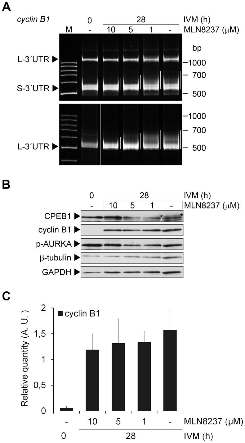
Figure 4. Cyclin B1 mRNA polyadenylation and cyclin B1 expression after AURKA inhibition.
(A) Polyadenylation of short (S-3′UTR) and long (L-3′UTR) forms of the cyclin B1 mRNA was examined by poly(A)-test in oocytes collected before and after 28 h of IVM in media supplemented with stated concentrations of MLN8237. The polyadenylation is highlighted by white lines next to each lane. (B) Oocytes collected before IVM and after 28 h of IVM in media supplemented with stated concentrations of MLN8237 were subjected to western blot analysis of CPEB1, cyclin B1 and phospho-AURKA using specific antibodies. β-tubulin and glyceraldehyde-3-phosphate dehydrogenase (GAPDH) were used as loading controls. The phosphorylated form of CPEB1 is marked (*). (C) The protein expression of cyclin B1 from six independent experiments was quantified using Quantity one software. The density of individual band was normalized to the total density of examined bands and to β-tubulin. The values represent the means ± SEM. No significant difference was detected between the control oocytes after 28 hours of IVM and the oocytes treated with different concentrations of MLN8237, P>0.05.

