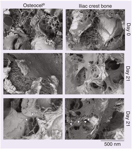
Figure 3. Environmental scanning electron microscopy of stromal cell abundance (Day 0, top panels) and in vitro scaffold colonization (Day 21, middle and bottom panels) in Osteocel® and control iliac crest bone.
White filled arrows show cell attachment points to bone, fat cells are indicated by dotted outline and black filled arrows.

