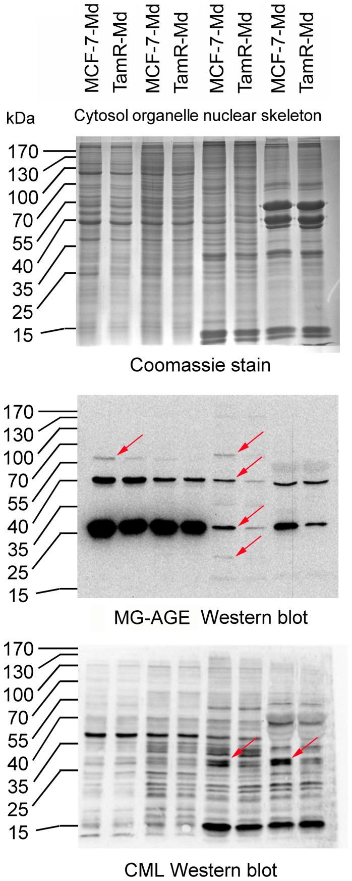
Figure 2. AGE accumulation in MCF-7 and TamR cells.
MCF-7-Md and TamR-Md cells were grown under standard conditions and harvested in the logarithmic phase. Cells were fractionated by a detergent based protocol and AGEs detected by Western blotting. Based on protein determination and Coomassie staining (A), equal amounts of protein were loaded for both cell lines and the AGEs MG-AGE (B) and CML (C) detected by Western analysis. Differences in the pattern of AGE-modified proteins are indicated by arrows.

